Yersinia
CASE 1
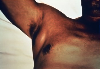
• A 35 year old Ghanaian engineer working on the construction of a new road in Botswana develops an enlarging, painful swelling in the right axilla over the past 2 days (shown below). The swelling is accompanied by fever, headache, and muscle aches.
• During the course of the road-building project, the patient slept on a sleeping bag on the ground under a canopy cover. He noticed some insect bites on the exposed parts of his body.
• When the patient is transported to the hospital, his temperature is 38.9 degrees and his blood pressure is 90/60 mmHg. The axillary lesion is exquisitely tender. The lesion is fluctuant (suggesting liquid content at the center), and there is early necrosis of the overlying skin.
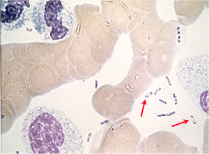
• The axillary mass is aspirated, and a Wayson stain is performed. Organisms with bipolar staining (like a "safety pin") are seen and are thought to be characteristic of Yersinia pestis. The patient was treated with ciprofloxacin and gentamicin and recovered in 2-3 weeks without any complications.
Questions:
1. What disease is caused by Yersinia pestis?
2. This is the bubonic form of the disease. Into what other forms might this disease evolve?
3. Was this infection acquired by an arthropod bite? If so, by what type arthropod?
4. Is this infection a zoonosis? What is the natural reservoir?
5. What is the difference between the sylvatic and urban cycle of this disease?
6. Can this patient's infection be transmitted to other humans? If so, how?
7. This pathogen constructs an elaborate extension from its surface to interact with host phagocytes (type III secretion system)? How does it work and what is its purpose?
CASE 2
• A 19 year old European woman develops fever and diarrhea with abdominal pain that migrates from the umbilicus to the right lower quadrant. Her white blood cell count is elevated to 16, 500/mm3. A pelvic examination is unremarkable.
• A diagnosis of appendicitis is made, and the patient is taken to the surgical theatre.
• At surgery, the appendix appears normal; however, there is a cluster of enlarged and inflammed mesenteric lymph nodes in the right paracolic area. One of the nodes is biopsied and cultured, and Yersinia enterocolitica is isolated from the culture one week later.
Questions:
1. How was this infection acquired?
2. In what ways is this infection similar to the one described in Case 1 above?